Deigram Of Outside Leg Muscles / Understanding The It Band Harvard Gazette / Three types of leg pain musculoskeletal.. Groin strain treatment rehabilitation exercises although there is often swelling oedema as a result of a groin strain this is often not visible to the eye groin strains are graded 1 2 or 3 depending on the extent of the injury groin muscle diagram diagram muscles in groin area male groin muscle diagram diagram muscles in groin area male anatomy groin human photo groin. This video identifies all muscles of the lower leg. The muscles that make up the quadriceps are the strongest and leanest of all muscles in the body. This muscle is one of the ones that help. Collectively referred to as the hip adductors, the groin muscles are.
This muscle runs along the outside of the back of your thigh and attaches to the top of the fibula (the smaller of the two bones of your lower leg). The forearm will be parallel to the lower leg. These muscles include the gluteus maximus muscle (the largest muscle in the body) and the hamstrings group, which consists of the biceps femoris, semimembranosus, and semitendinosus muscles. Anterior compartment, also known as the extensor compartment; Posterior compartment, also known as the flexor compartment;
3 from Calf muscle anatomy, calf muscle picture anatomy, lateral calf muscles, lower leg muscle anatomy, medial calf muscles, what are the two calf muscles called, what are your calf muscles called, where is your calf muscle, human muscles, calf muscle anatomy, calf muscle picture anatomy, lateral calf. The hip muscles work together to carry out 4 different types of movement: The muscles of the lower leg, called simply the leg by anatomists, largely move the foot and toes. Groin muscles help support the hip joint. It contains the peroneus longus and peroneus brevis muscles. On the medial edge of the posterior thigh is the gracilis muscle. This type of leg pain is related to muscles or skeletal system. Anterior compartment, also known as the extensor compartment;
Medial compartment, also known as adductor compartment;
On the medial edge of the posterior thigh is the gracilis muscle. This group includes the adductor magnus, adductor longus, and adductor brevis muscles, as well as the pectineus and gracilis. Musculoskeletal leg pain is the most common form. This video identifies all muscles of the lower leg. These four muscles at the front of the thigh are the major extensors (help to extend the leg straight) of the knee. The calf muscle, on the back of the lower leg, is actually made up of two muscles: Leg pains can happen for a variety of reasons. The hip muscles work together to carry out 4 different types of movement: Muscle diagrams of major muscles exercised in below is an image of the outside of a normal healthy human heart diagram. The gastrocnemius is the larger calf muscle, forming the bulge visible beneath the skin. The following diagram illustrates the actions of the terms adduction, abduction, flexion and extension at the different joints. This muscle is one of the ones that help. They allow you to move and provide support for your upper body.
Tendons in upper leg / upper leg muscles diagram quizlet. The forearm will be parallel to the lower leg. However, many reflex pathways are also active in the legs and foot. Conditions seen in this category are muscle strain, ligament and tendon strain. 18 photos of the lower leg muscles diagram.
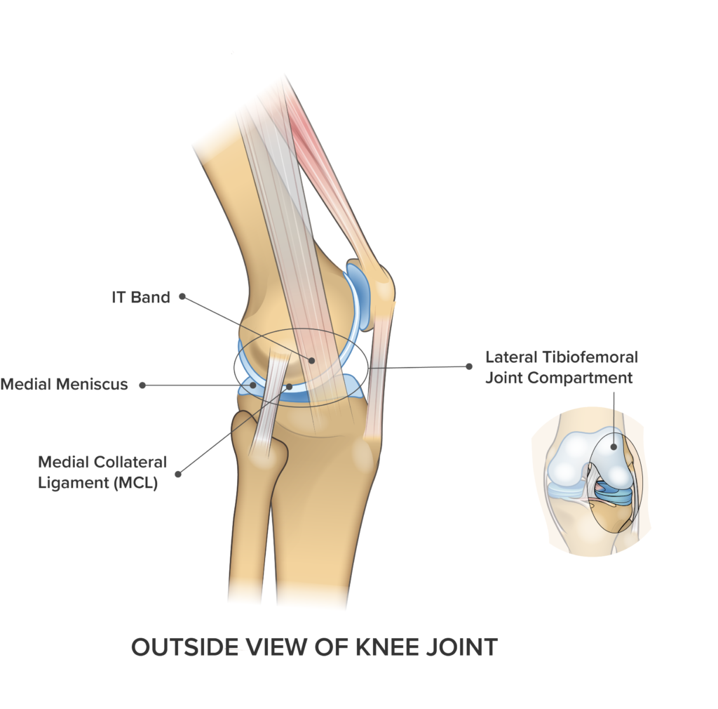
Knee Pain On The Outside Of Your Joint Five Reasons Why Spring Loaded Technology from springloadedtechnology.com The hip muscles work together to carry out 4 different types of movement: Your quadricep muscles, also known as quads, consist of four muscles that compose the front of your leg; Tendons can be small, like the delicate, tiny bands in the hands, or large, like the heavy, ropelike cords that anchor the calf or thigh muscles. This anatomy chart is a great example of beauty and function in one, as it is pleasing to look… Medial compartment, also known as adductor compartment; The muscles in the hip are responsible for the movement of the hip and, by proxy, the leg. The muscles work together to enable movement and keep the hip in alignment. Human muscle system, the muscles of the human body that work the skeletal system, that are under voluntary control, and that are concerned with movement.
These muscles include the gluteus maximus muscle (the largest muscle in the body) and the hamstrings group, which consists of the biceps femoris, semimembranosus, and semitendinosus muscles.
Add a frame to any. It gets its blood flow from the arteries in the tiberial artery. Your quadricep muscles, also known as quads, consist of four muscles that compose the front of your leg; The following diagram illustrates the actions of the terms adduction, abduction, flexion and extension at the different joints. Anterior compartment leg muscles the anterior compartment of the leg comprises four muscles. The forearm will be parallel to the lower leg. Tendons can be small, like the delicate, tiny bands in the hands, or large, like the heavy, ropelike cords that anchor the calf or thigh muscles. This group includes the adductor magnus, adductor longus, and adductor brevis muscles, as well as the pectineus and gracilis. Collectively referred to as the hip adductors, the groin muscles are. Anterior muscles of the lower leg and their functions. On the outside of the thigh, this is the largest of. This is why you have to indicate which biceps you are taking about when discussing one or other of these muscles. To feel these muscles contract, place your hand on the outside of your shin and turn your foot out.
The gastrocnemius is the larger calf muscle, forming the bulge visible beneath the skin. Groin strain treatment rehabilitation exercises although there is often swelling oedema as a result of a groin strain this is often not visible to the eye groin strains are graded 1 2 or 3 depending on the extent of the injury groin muscle diagram diagram muscles in groin area male groin muscle diagram diagram muscles in groin area male anatomy groin human photo groin. This video identifies all muscles of the lower leg. Extension, flexion, adduction, and abduction. However, all leg pains are categorized into three major types.
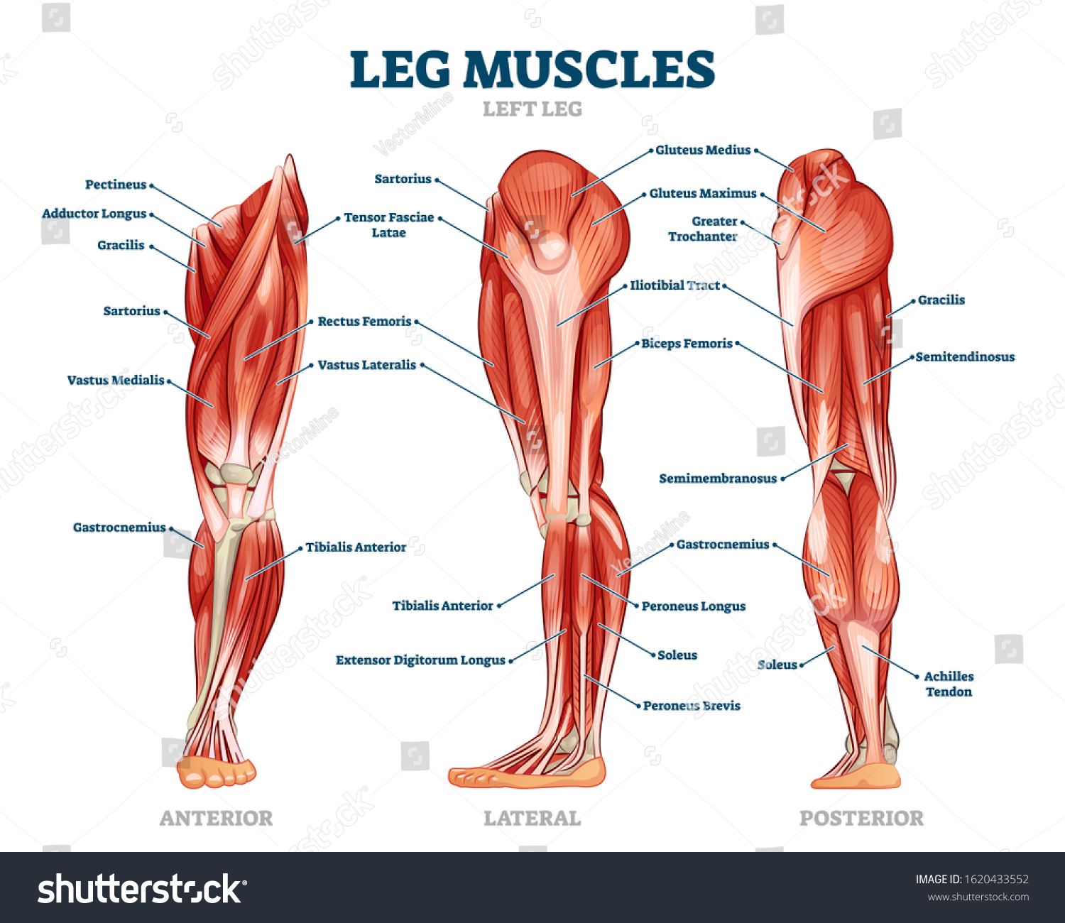
Leg Muscle Anatomical Structure Labeled Front Stock Vector Royalty Free 1620433552 from image.shutterstock.com This is why you have to indicate which biceps you are taking about when discussing one or other of these muscles. It is also visible on the medial edge of the thigh from the anterior. Anterior muscles of the lower leg and their functions. These four muscles at the front of the thigh are the major extensors (help to extend the leg straight) of the knee. The muscles work together to enable movement and keep the hip in alignment. We'll break down the anatomy and function of the upper leg, knee, lower leg. Conditions seen in this category are muscle strain, ligament and tendon strain. Your quadricep muscles, also known as quads, consist of four muscles that compose the front of your leg;
When tendons become inflamed, irritated or suffer microscopic tears, the condition is called tendonitis.
The hip muscles work together to carry out 4 different types of movement: Conditions seen in this category are muscle strain, ligament and tendon strain. The anterior compartment of the leg acts to dorsiflex and invert the foot through the ankle joint. The anterior is located in the front portion of the leg. Leg pains can happen for a variety of reasons. Human muscle system, the muscles of the human body that work the skeletal system, that are under voluntary control, and that are concerned with movement. The lateral compartment is along the outside of the lower leg. On the medial edge of the posterior thigh is the gracilis muscle. Included are several layered views of the back muscles, the dorsal muscles, subclavius muscles, rhomboideus major and minor muscles, deltoid muscles and many more. These four muscles at the front of the thigh are the major extensors (help to extend the leg straight) of the knee. The leg muscles diagram, will point out if the issue is with any tissue or with the bone. A muscle strain is a stretch or tear of muscle fibers. These muscles pull the toes and feet outward.


0 Komentar